Arthritis MRI Symptom Checker
Select Your Symptoms
Click on the symptoms you are currently experiencing. The tool will analyze your selection against common arthritis patterns.
Analysis Results
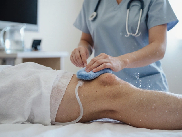
You wake up with a stiff knee or a swollen finger. It’s been bothering you for weeks, maybe months. Your doctor orders an MRI (Magnetic Resonance Imaging) scan. But here is the question burning in your mind: Will this machine actually show me if I have arthritis? The short answer is yes. In fact, an MRI is one of the most powerful tools we have for seeing arthritis long before it shows up on other tests.
But it’s not that simple. An MRI doesn’t just say "yes" or "no." It reveals the story of what is happening inside your joints. It can spot inflammation, cartilage wear, and even tiny bone changes that X-rays miss completely. Understanding what an MRI can and cannot see helps you interpret your results and talk smarter to your orthopedic specialist.
Why MRIs Are Different From X-Rays
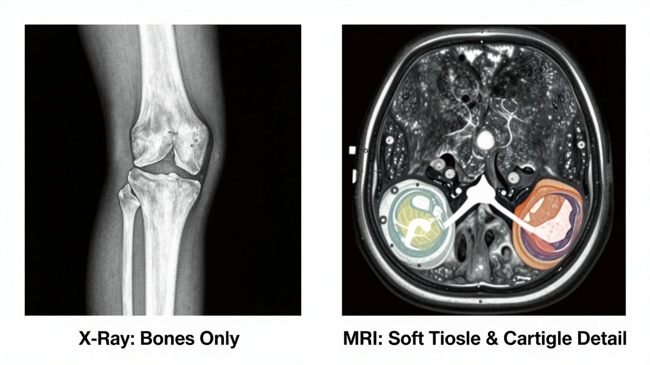
To understand why an MRI is so good at spotting arthritis, you first need to know how it differs from the standard X-ray. Most people think of X-rays as the go-to for bone issues. And they are great for seeing fractures or advanced bone spurs. But X-rays only show dense material like bone. They cannot see soft tissue.
Arthritis isn’t just about bones grinding together. It involves the cartilage, the synovium (the lining of the joint), ligaments, and tendons. An X-ray might show that two bones are close together because the cartilage has worn down, but it won’t show the cartilage itself. It’s like looking at a car engine through a thick fog-you can see the block, but you can’t see the belts or hoses failing.
An MRI uses strong magnetic fields and radio waves to create detailed images of both hard and soft tissues. This means it can visualize the actual thickness of your cartilage, the health of your meniscus in the knee, and whether there is fluid buildup indicating active inflammation. For conditions like rheumatoid arthritis, where the immune system attacks the joint lining, an MRI is often the only way to catch the disease in its earliest stages.
What Exactly Does an MRI Show?
When a radiologist looks at an MRI for signs of arthritis, they are hunting for specific clues. These aren’t just vague shadows; they are distinct structural changes that point directly to joint degeneration or autoimmune attack.
- Cartilage Loss: Healthy cartilage appears as a smooth, dark line covering the ends of bones. If it’s thinning, fraying, or missing entirely, the MRI shows it clearly. This is a hallmark of osteoarthritis.
- Synovitis: This is inflammation of the joint lining. On an MRI, especially with contrast dye, inflamed tissue lights up bright white. This is crucial for diagnosing inflammatory arthritis types.
- Bone Marrow Edema: This sounds scary, but it simply means there is fluid inside the bone due to stress or inflammation. It’s a sign that the joint is under significant pressure or attack.
- Erosions: In aggressive forms of arthritis, the bone itself gets eaten away. An MRI can detect these tiny holes or rough patches on the bone surface before they become large enough to see on an X-ray.
- Ligament and Tendon Damage: Arthritis often weakens the structures holding the joint together. Tears or stretches in these tissues contribute to instability and pain.
Osteoarthritis vs. Rheumatoid Arthritis on Scans
Not all arthritis is the same. The type of arthritis you have dictates what the MRI will look like. Let’s break down the two most common types.
| Feature | Osteoarthritis (OA) | Rheumatoid Arthritis (RA) |
|---|---|---|
| Primary Cause | Wear and tear / Mechanical stress | Autoimmune system attacking joints |
| Cartilage Appearance | Focal loss, fraying, cracks | Diffuse thinning, rapid degradation |
| Inflammation Level | Mild to moderate (secondary) | High (primary driver of damage) |
| Bone Changes | Bone spurs (osteophytes), sclerosis | Erosions, pannus formation |
| Joints Affected | Knees, hips, spine, hands | Hands, wrists, feet (often symmetrical) |
In osteoarthritis, the MRI tells a story of mechanical failure. You’ll see uneven gaps where cartilage used to be, often accompanied by bone spurs trying to stabilize the joint. It’s localized. If your right knee hurts, the left one might look perfectly fine.
In rheumatoid arthritis, the story is systemic. The MRI shows widespread inflammation. You’ll see the synovium thickening and invading the bone. This is called pannus formation. It’s aggressive. RA often affects both sides of the body symmetrically-if your right wrist is inflamed, your left likely is too.
Can MRI Detect Early-Stage Arthritis?
This is the biggest advantage of MRI technology. Traditional X-rays often come back "normal" in patients who are in significant pain. Why? Because X-rays only show damage after about 30-50% of the cartilage is gone. By then, the joint is already compromised.
An MRI can detect pre-clinical arthritis. It can identify micro-fractures in the bone plate beneath the cartilage, subtle changes in cartilage composition, and early synovial inflammation. For athletes or young adults with unexplained joint pain, an MRI can confirm that arthritis is starting years before it becomes a disability. Catching it early allows for interventions like physical therapy, lifestyle changes, or targeted medications that can slow progression significantly.
The Role of Contrast Dye
Sometimes, your doctor will order an MRI with contrast. This involves injecting a gadolinium-based dye into your vein before the scan. Why do this?
Contrast dye highlights blood flow and inflammation. Active arthritis is vascular-it has increased blood supply to the inflamed areas. Without contrast, chronic scar tissue and active inflammation can look similar. With contrast, the active inflammation lights up brightly. This distinction is vital for treating inflammatory arthritis. If the inflammation is active, anti-inflammatory drugs or biologics might work. If it’s just scar tissue, those drugs won’t help much, and surgery or physical therapy might be better options.
Note: If you have kidney issues or a known allergy to gadolinium, tell your doctor immediately. There are alternatives, but safety comes first.
Limitations of MRI for Arthritis
While powerful, MRI isn’t magic. It has limitations you should be aware of.
Cost and Time: MRIs are expensive and take longer than X-rays. A full knee MRI can take 30-45 minutes. You have to lie still in a confined space. For people with claustrophobia, this can be challenging (though open MRIs exist).
Over-diagnosis: Sometimes, an MRI shows "abnormalities" that don’t cause pain. Many healthy people over 40 have mild cartilage wear or small bone spurs on their MRI scans but feel no pain at all. Doctors must correlate the MRI findings with your physical symptoms. Just because the scan looks bad doesn’t always mean you need surgery.
Metal Implants: If you have certain metal implants, pacemakers, or cochlear implants, you may not be able to undergo an MRI. The magnetic field can interfere with these devices. Always disclose any medical hardware to the technician.
What Happens After the Scan?
Once the MRI is done, a radiologist reads the images and sends a report to your doctor. This report will describe the grade of cartilage loss, the presence of effusion (fluid), and any bone marrow lesions. Your orthopedic specialist will then combine this data with your physical exam-checking for range of motion, swelling, and tenderness-to create a treatment plan.
If the MRI shows early-stage osteoarthritis, the focus might be on weight management, strengthening exercises, and possibly corticosteroid injections. If it shows advanced rheumatoid arthritis with erosions, you might be referred to a rheumatologist for disease-modifying antirheumatic drugs (DMARDs). In severe cases where bone-on-bone contact is evident and pain is unmanageable, joint replacement surgery might be discussed.
Preparing for Your MRI
To get the clearest possible images, preparation matters. Wear loose, comfortable clothing without metal zippers, hooks, or underwires. You may be asked to change into a hospital gown. Remove all jewelry, watches, and hearing aids. If you have tattoos, mention them; some inks contain metal that can heat up or distort images, though this is rare with modern ink.
If you are anxious, ask if sedation is an option. Some centers offer mild sedation for claustrophobic patients. Also, remember to breathe normally during the scan. Holding your breath can cause motion artifacts that blur the image.
Is an MRI painful?
No, the MRI procedure itself is painless. You lie on a table that slides into a tube-like machine. You might hear loud knocking or buzzing noises as the magnets switch on and off, which is normal. Earplugs or headphones are provided to reduce noise. The main discomfort is lying still for 30-60 minutes.
How soon will I get my MRI results?
Typically, results are available within 24 to 72 hours. The radiologist writes a detailed report, which is sent to your referring physician. Your doctor will review the images and report with you during a follow-up appointment to explain what the findings mean for your treatment.
Can an MRI rule out arthritis completely?
An MRI is highly sensitive, but it’s part of a bigger puzzle. If your MRI is normal but you have persistent joint pain, swelling, and stiffness, your doctor may still suspect inflammatory arthritis. Blood tests (like RF or Anti-CCP) and clinical history are also critical. A normal MRI makes advanced osteoarthritis unlikely, but it doesn’t rule out early inflammatory conditions.
Do I need an MRI if my X-ray was normal?
Yes, sometimes. If your X-ray is normal but you have significant pain, limited mobility, or suspected soft tissue injury (like a meniscus tear or ligament sprain), an MRI provides the detail needed. It checks for soft tissue problems and early cartilage damage that X-rays miss.
Are there risks associated with MRI scans?
MRI does not use ionizing radiation like X-rays or CT scans, making it safer in that regard. The main risks involve the strong magnetic field, which can affect metal implants or pacemakers. Contrast dye carries a small risk of allergic reaction or kidney issues in people with pre-existing renal disease. Always discuss your medical history with your provider.