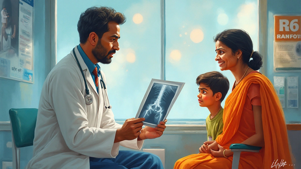
Walk into any ER with a broken arm, and you might hear a group of doctors tossing around phrases like "the 4 A’s." For most people, that sounds like hospital code—or maybe a weird school grading system. But for orthopedic surgeons, those A’s are the backbone (pun intended) of bone and joint care. These four critical steps—Alignment, Apposition, Apparatus, and Activity—guide everything from a simple finger fracture to hip replacements in athletes and accident victims. And while it may look straightforward on a textbook chart, applying all four perfectly in the chaos of a trauma center is a whole other game.
So, what do these A's actually mean? And why should you care if you’ve ever tripped over a scooter or taken a tumble off your cycle? Sometimes, knowing the basics could mean faster recovery, less pain, and a better outcome. Let’s break it down and really look at how the world of orthopedics leans on these pillars.
Alignment: Getting Things Straight, Literally
Imagine if your broken bone healed crooked. Not fun, right? That’s where alignment comes in—making sure the ends of the bone or joint line up as naturally as possible. If a bone is misaligned when it heals, things like movement, function, and even cosmetics can take a hit. According to the British Orthopaedic Association, perfect or near-perfect alignment sharply improves the chances of regaining full movement after a break. Surgeons often use physical manipulation, special beds, or even weights (like traction) to pull things into place.
This isn’t a "close enough" business. Even a few degrees off can mess up the way you walk or run. In young kids, bones can sometimes "remodel" themselves over time if the alignment is slightly off, but adults don’t get that lucky. That’s why your doctor might push, pull, or twist a limb—even if it hurts short-term. The payoff is living pain-free later on.
If the break is more complicated, the team may bring out X-rays or even 3D imaging. Fun fact: Hospitals in India have been leading the way in using AI to check alignment in digital scans, helping docs spot tiny misalignments right after a cast goes on. This isn’t just about looking good on the outside—a bad alignment can wear out joints early or cause arthritis years down the line.
Apposition: Keeping the Pieces Close
Picture a puzzle with some pieces hovering across the table instead of locked together. That’s poor apposition. In orthopedics, apposition means the bone ends are touching as much as possible—giving them a chance to heal like they did before the break. The classic tip surgeons give: "Contact heals bone." It’s about creating a stable environment where bone cells rebuild efficiently.
Doctors check apposition by both touch and imaging. A good sense of touch is part instinct, part hardcore training—surgeons actually practice with models to learn what a well-matched bone feels like. In tech-savvy hospitals, real-time portable X-ray machines (called image intensifiers) let teams double-check their work. In rural areas, where fancy hardware isn’t always available, a seasoned orthopedic doctor’s hands are sometimes the best tool.
Be aware, poor apposition slows down healing. There’s a higher risk of "non-union," where the bone just refuses to knit back together. And then you’re dealing with more surgeries or maybe even long-term disability. That’s why sports medicine clinics are obsessed with getting this step right—athletes need bones to heal strong and fast, not weak or wobbly.

Apparatus: Using the Right Tools
This part sounds simple—set the bone and use something to keep it in place. Reality check: picking the right apparatus can mean the difference between a full recovery and a lifetime of problems. Apparatus includes casts, splints, braces, wires, plates, screws, or external fixators that stabilize the bone so it can heal undisturbed.
Here’s a cool tidbit: In one big teaching hospital, surgeons used an external fixator on a complicated leg fracture and cut the healing time by 30% compared to the old cast-and-bed-rest routine. Another example—kids with forearm fractures often heal faster with newer, lightweight fiberglass casts, so they can get moving again without risking more damage. What’s hot in 2025? 3D-printed casts fitted like a glove and water-resistant, so no more smelly, soggy arms after a shower.
The apparatus also needs regular checks. Even the best cast can cause sores, cuts, or numbness if it’s too tight or slips out of position. Some hospitals give patients a schedule for checking their skin color, temperature, or movement—those are early warning signs that it’s time to ring the clinic. Doctors also weigh the pros and cons of "rigid" (immovable) apparatus versus "functional" ones that allow early movement. Elderly patients often do better with braces that let them move a bit while healing—less risk of blood clots and muscle loss.
| Apparatus Type | Best For | Advantages | Common Issues |
|---|---|---|---|
| Traditional Cast | Simple fractures | Cheap, available everywhere | Heavy, can’t get wet, itchy |
| 3D-Printed Cast | Complicated or awkward fractures | Light, custom fit, water-resistant | Expensive, needs imaging & expertise |
| External Fixator | Severe trauma, open wounds | Fixes complex cases, adjustable | Bulky, risk of infection at pin sites |
| Surgical Plates & Screws | Fractures near joints or in adults | Stable, allows earlier activity | Surgical risks, possible removal later |
Activity: Moving at the Right Time
This last “A” can feel like a juggling act—push too soon, and you risk breaking the repair; wait too long, and you face muscle waste and joint stiffness. Hospitals now focus on "early mobilization," which means getting people moving much sooner than a decade ago. Back in the day, bedrest after a fracture could last weeks. Today, research from the Indian Orthopaedic Association shows that early movement drops complications like blood clots and joint contractures by up to 40%.
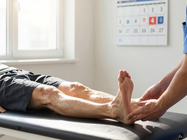
This isn’t about running before you can walk. It’s about careful, guided movements: a few fingers at first, maybe gentle stretches. For more severe injuries—like hip replacements—patients often stand up on day one with a walker. The key is gradual load. Too little pressure and bones stay weak. Too much, and you re-break what just healed. Physical therapists create step-by-step plans, usually adjusting weekly based on pain, swelling, and X-ray progress.
A solid tip: ask your care team for a home exercise routine. Patients who stick to daily moves, like ankle pumps or gentle grip squeezes, recover faster and get back to normal activities—school, work, or sports. Some clinics even give out digital trackers that buzz every hour as a reminder. And don’t ignore warning signs—pain that won’t quit, tingling, or swelling could mean something’s off.
Here’s a wild stat: In a Mumbai study last year, 85% of patients who kept moving within two days of a wrist fracture had a stronger grip three months later than those who waited a week. That’s a huge difference in daily life, from opening jars to typing up work emails. So yes, activity is more than just getting by—it’s about coming out stronger.
