Bone & Joint Diagnostic Finder
Select your primary symptom or concern below to find the recommended first-line diagnostic test, estimated cost in India, and why it is chosen.
Test Name
CategoryWhy this test?
Reasoning goes here.
Important Notes
Additional context.
You wake up with a stiff knee or a nagging back pain that won't quit. You visit an orthopedic specialist, and they hand you a slip for a scan. But which one? An X-ray? An MRI? A CT scan? It’s confusing, expensive, and frankly, scary if you don’t know what you’re looking at.
Here is the truth: there is no single "best" test for everyone. The right diagnostic tool depends entirely on what tissue is hurting-your hard bone, your soft cartilage, your ligaments, or your nerves. Using the wrong test is like using a hammer to fix a watch; it might make noise, but it won’t solve the problem. This guide breaks down exactly which imaging technique works best for specific bone and joint issues, so you can ask the right questions and avoid unnecessary costs.
The Quick Verdict: Which Test Do You Need?
| Symptom / Issue | Best First-Line Test | Why? | Cost Estimate (India) |
|---|---|---|---|
| Bone Fracture | X-Ray | Fast, cheap, shows broken bones clearly. | ₹500 - ₹1,500 |
| Ligament/Tendon Tear | MRI | Shows soft tissues (ACL, meniscus) that X-rays miss. | ₹4,000 - ₹8,000 |
| Arthritis/Joint Degeneration | X-Ray + Clinical Exam | Shows joint space narrowing and bone spurs. | ₹500 - ₹1,500 |
| Complex Bone Tumor | CT Scan | 3D view of complex bone structures. | ₹3,000 - ₹6,000 |
| Osteoporosis | DEXA Scan | Measures bone mineral density specifically. | ₹2,500 - ₹4,500 |
X-Rays: The Workhorse for Hard Tissues
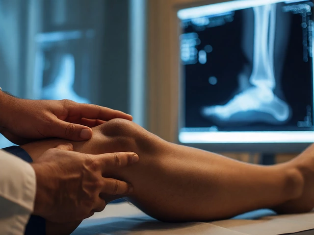
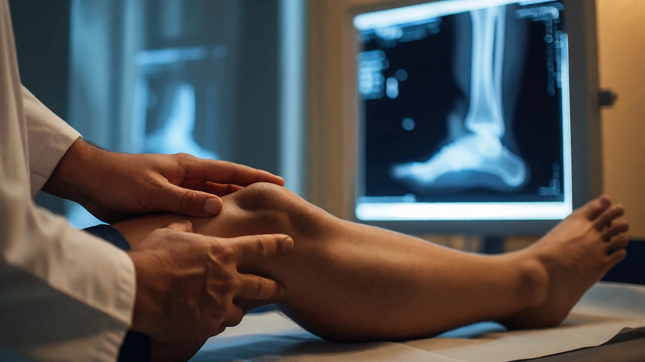
If you fall and twist your ankle, the doctor will almost always order an X-ray first. Why? Because it’s fast, widely available, and incredibly good at showing dense materials like calcium in your bones. If you have a fracture, a dislocation, or significant arthritis, an X-ray will likely spot it immediately.
But here is the catch: X-rays are terrible at showing soft tissue. If you tore your ACL (a ligament in your knee) or damaged your meniscus (the cushion in your knee), an X-ray might look completely normal even though you are in severe pain. This is why patients often get frustrated when their "normal" X-ray doesn't explain their agony. Remember, a normal X-ray only means your bones aren't broken; it says nothing about your muscles, tendons, or nerves.
Pro Tip: Always keep your old X-rays. Comparing a current image with one from five years ago helps doctors see how quickly arthritis is progressing or if a previous injury has healed incorrectly.
MRI: The Gold Standard for Soft Tissue and Nerves
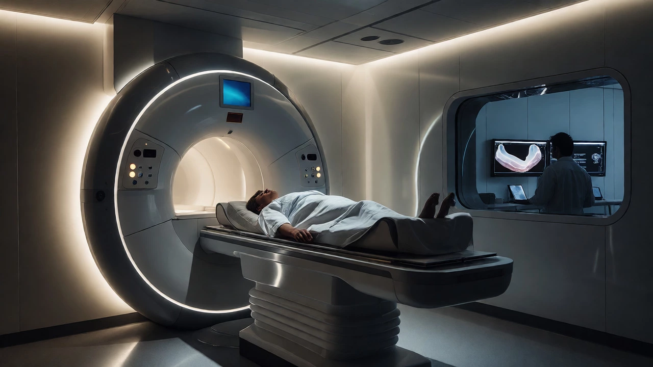
When the problem isn't in the bone, but around it, you need an MRI (Magnetic Resonance Imaging). Unlike X-rays, MRIs use powerful magnets and radio waves, not radiation. This allows them to create detailed cross-sectional images of every structure in your body.
MRI is the go-to choice for:
- Knee Injuries: Torn ACL, PCL, or meniscus tears.
- Spine Issues: Herniated discs pressing on nerves (sciatica).
- Shoulder Pain: Rotator cuff tears or labral damage.
- Early Stress Fractures: Sometimes a hairline fracture doesn't show on X-ray until weeks later, but an MRI catches it instantly.
The downside? It takes longer (30-60 minutes), it's loud, and it costs significantly more. Also, if you have certain metal implants, pacemakers, or claustrophobia, an MRI might be tricky or impossible. However, for anything involving pain without a visible break, this is usually the most valuable test you can buy.
CT Scans: Seeing the Complex Geometry
A CT Scan (Computed Tomography) is essentially a super-charged X-ray. It takes hundreds of X-ray images from different angles and uses a computer to stitch them into 3D slices. While an X-ray gives you a flat shadow, a CT gives you a topographic map of your skeleton.
Doctors rarely use CT scans for simple sprains. They reserve them for complex scenarios:
- Complex Fractures: Like those in the wrist, spine, or pelvis where alignment matters for surgery.
- Bone Tumors: To see the exact size and shape of a growth.
- Pre-Surgical Planning: Surgeons need 3D models to plan hardware placement.
Be aware that CT scans involve higher radiation doses than standard X-rays. So, unless your doctor suspects something complex that an X-ray missed, you probably don't need one right away.
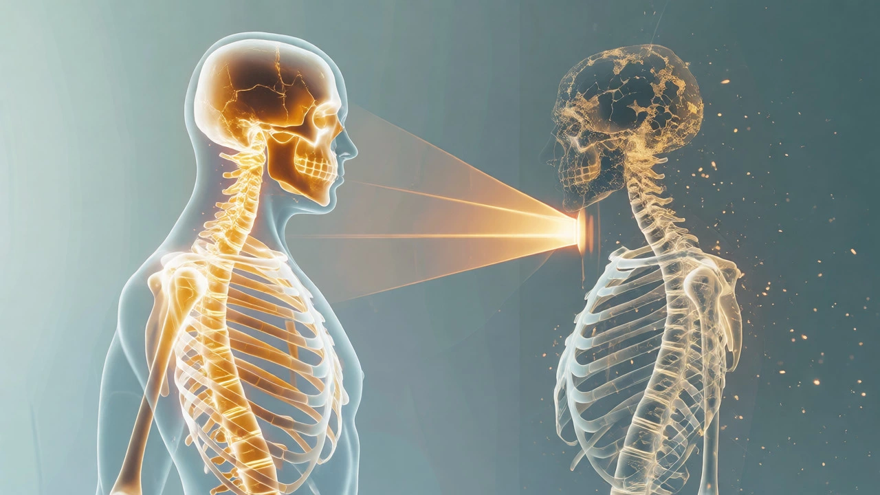
Bone Density Tests: Checking for Osteoporosis
If you are over 50, especially if you are post-menopausal, or if you have had multiple fractures, your doctor might worry about bone quality rather than just bone structure. That’s where the DEXA Scan (Dual-Energy X-ray Absorptiometry) comes in.
This isn't a diagnostic test for acute pain. It’s a screening tool. It measures how much calcium and other minerals are packed into your bones. Low density means brittle bones (osteoporosis), which puts you at risk for future breaks. It’s quick, painless, and involves very low radiation. If you’ve lost height recently or stooped forward, ask for this test. It’s preventive medicine, not reactive treatment.
Ultrasound: The Dynamic Option
Many people forget that Ultrasound is a legitimate orthopedic tool. It uses sound waves to create real-time images. This is fantastic for superficial joints like shoulders, elbows, wrists, and ankles.
Why choose ultrasound? Because it’s dynamic. With an MRI, you lie still. With an ultrasound, the doctor can move your arm while watching the screen. They can see if a tendon snaps or slips out of place when you move. It’s also great for guiding injections, ensuring the needle hits the exact spot. Plus, it’s cheaper and faster than an MRI. For things like bursitis, tendonitis, or ganglion cysts, ultrasound is often better than an MRI because it captures movement.
How to Choose: A Decision Guide
Feeling overwhelmed? Use this simple logic flow before you book an appointment:
- Did you have trauma (fall/accident)? Start with an X-ray. If negative but pain persists, consider MRI.
- Is the pain sharp and localized to a specific tendon? Try Ultrasound first. It’s cheaper and sees surface issues well.
- Do you have numbness, tingling, or weakness? This suggests nerve involvement. Go straight for an MRI (especially for spine issues).
- Are you older with gradual stiffness? Start with X-rays to check for osteoarthritis. Add DEXA if fracture risk is high.
- Is the diagnosis unclear after initial tests? That’s when advanced options like CT or specialized MRIs come into play.
Pitfalls to Avoid
Don't self-prescribe imaging. Walking into a radiology center and asking for a full-body MRI is a waste of money. Radiologists read what the referring doctor asks for. If you don't have a clear clinical question, the report might be vague or misleading.
Also, beware of "incidental findings." As imaging gets better, we find tiny abnormalities that never caused symptoms. Treating these can lead to unnecessary surgeries. Only act on imaging results if they match your physical symptoms. If your knee hurts on the left side, but the MRI shows a minor tear on the right, the tear might not be the culprit.
Which is cheaper, X-ray or MRI?
X-rays are significantly cheaper, typically costing between ₹500 and ₹1,500 in India. MRIs are more expensive, ranging from ₹4,000 to ₹8,000 depending on the city and hospital. However, if an X-ray misses a soft tissue injury, you may end up paying for both, making the initial MRI cost-effective in some cases.
Can an X-ray detect arthritis?
Yes, X-rays are excellent for detecting moderate to severe osteoarthritis. They show joint space narrowing, bone spurs (osteophytes), and bone deformities. However, they cannot detect early-stage inflammation or cartilage loss before it affects the bone, which is where MRI or clinical exams become necessary.
Is an MRI safe?
MRIs do not use ionizing radiation, making them safer than CT scans or X-rays regarding radiation exposure. However, the strong magnetic field can interfere with pacemakers, cochlear implants, or certain metallic clips. Always inform your technician about any metal implants or devices before entering the machine.
What is a bone scan and when is it used?
A bone scan involves injecting a small amount of radioactive tracer into your bloodstream. It highlights areas of high bone activity. It is rarely used for common joint pain but is valuable for detecting metastatic cancer spreading to bones, occult stress fractures, or infections (osteomyelitis) that other tests miss.
How long does it take to get results?
X-ray reports are usually available within 15-30 minutes. Ultrasound reports are often given immediately by the sonographer. MRI and CT scans require a radiologist to review the images, so written reports typically take 24 to 48 hours, though emergency cases are prioritized.